

Urinary tract infections (UTIs) are common in young children. It has been estimated that up to 7% of girls and 2% of boys will have a UTI in the first 6 years of life. Many of these children with recurrent UTIs have VUR. Children with VUR are at risk of renal scarring. This scarring can cause serious sequelae as these children grow into adulthood, including renal hypertension, proteinuria, and end-stage renal disease. The negative health impacts of VUR can be successfully ameliorated by prompt diagnosis, allowing for early management with antibiotic prophylaxis to prevent UTIs, and surgical interventions in more severe cases
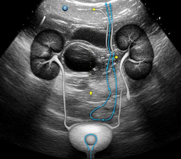
Currently, recommendations are that children with febrile or recurrent UTIs undergo diagnostic imaging to evaluate for the presence of VUR. Three imaging modalities are currently available for VUR detection: voiding cystourethrography (VCUG), direct radionuclide cystography (DRNC), and contrast-enhanced voiding ultrasonography (CE-VUS). Many children, once diagnosed with VUR, may require serial imaging to guide treatment; therefore, the safety and cost of the imaging modality are important considerations. CE-VUS, widely used for decades primarily in Europe, was recently introduced in the United States. CE-VUS has high diagnostic accuracy in detecting reflux and does not expose children to ionizing radiation
After completing this activity, participants should be better able to:
- Describe indications and accepted uses of CEUS of the kidneys, bladder, and urinary tract
- Summarize practice guidelines, recommendations, and clinical trials demonstrating the clinical utility of CEUS
- Review physicochemical, acoustic, and pharmacodynamics/pharmacokinetic characteristics of ultrasound contrast
agents (UCAs)
This activity is designed for pediatric radiologists, pediatric urologists, sonographers, nurses, and other healthcare providers involved with imaging of the urinary tract in pediatric patients with known or suspected vesicoureteric reflux (VUR) to help them better understand the indications, applications, and potential benefits of utilizing contrast-enhanced ultrasound (CEUS) for detecting known or suspected diseases and abnormalities of the kidneys, bladder, and urinary tract.
This SDMS CME activity has been planned and submitted for approval of 1.00 hour of SDMS CME Credit Category AB by the Society of Diagnostic Medical Sonographers (SDMS).
Release Date: 7/14/25 | Expires 7/14/2026
Faculty and Planner Disclosures:
As an accredited provider of continuing medical education, it is the policy of Northwest Imaging Forums, Inc. (NWIF) to ensure balance, independence, objectivity, and scientific rigor in all of its webinar activities. In accordance with this policy, faculty and planners must disclose any financial relationships with ineligible companies. Additionally, in the event a relevant financial relationship does exist, it is the policy of NWIF to ensure that the relevant financial relationship is mitigated in order to ensure the integrity of the SDMS CME activity. The planner has nothing to disclose.
Richard G. Barr, MD, PhD
Professor of Radiology
Northeast Ohio Medical University
Director, Southwoods Imaging
Department of Radiology
Surgical Hospital at Southwoods
Youngstown, Ohio

Aikaterini Ntoulia, MD, PhD
Department of Radiology
The Children’s Hospital of Philadelphia
Philadelphia, Pennsylvania