


Open to access this content
MRI CME
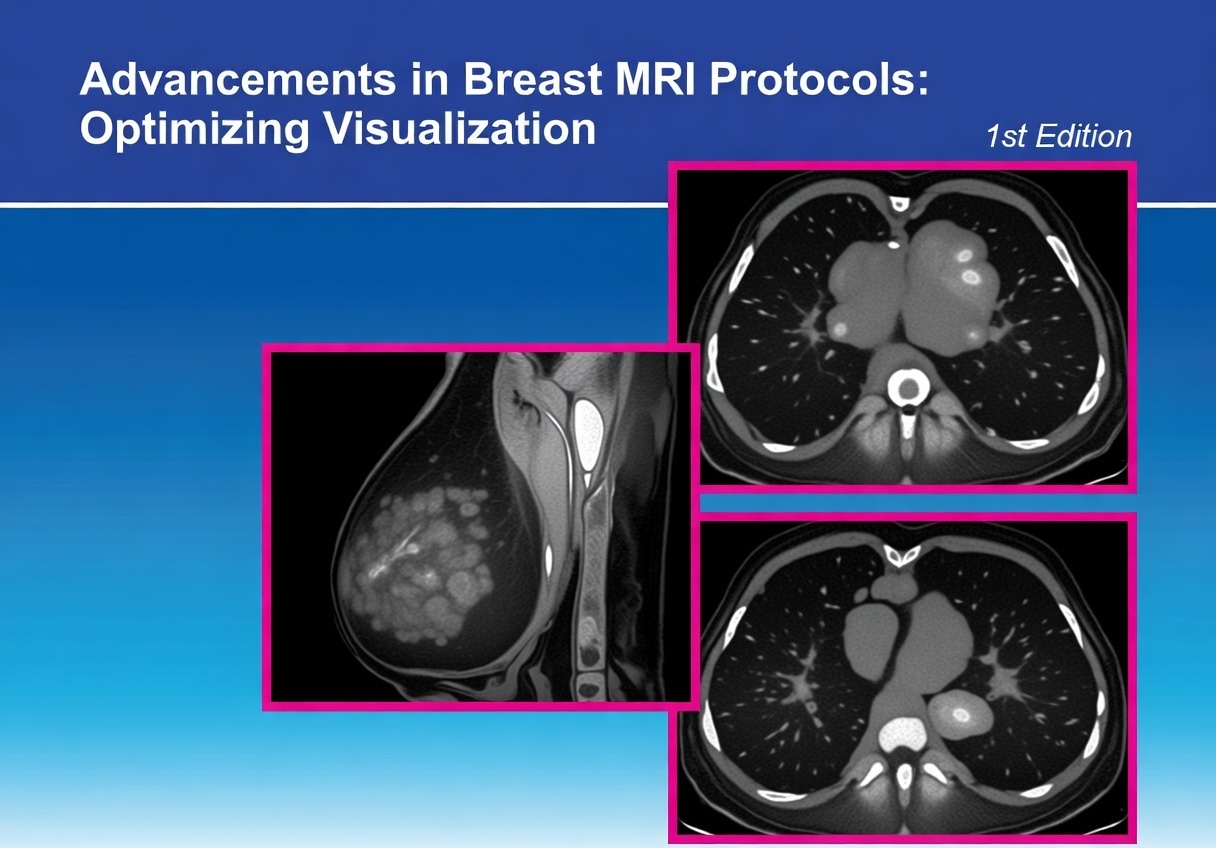
Advancements in Breast MRI Protocols: Optimizing Visualization – CME
Open to access this content
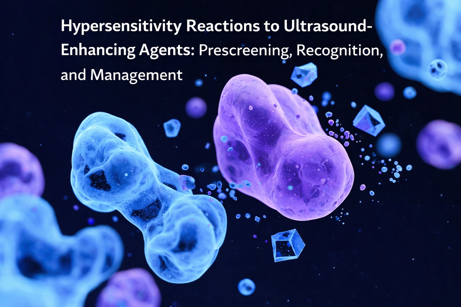
Hypersensitivity Reactions to Ultrasound Enhancing Agents: Prescreening, Recognition, and Management
Open to access this content

2026 – Level 2 MR Safety for MR Radiologists
Open to access this content

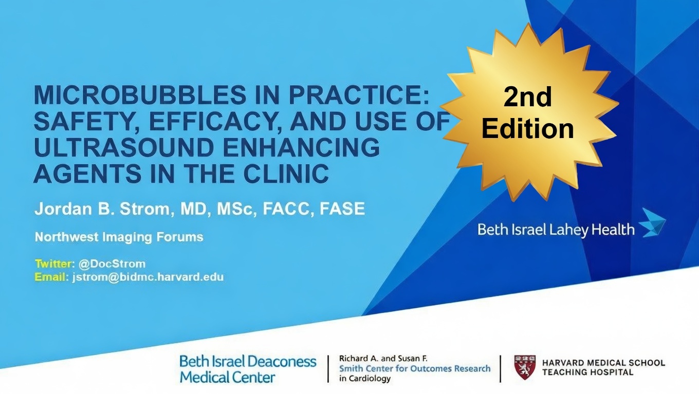
Use and Effectiveness of Contrast Enhanced Ultrasound (CEUS) for Echocardiography – CME
Open to access this content

The Safe and Effective Use of Ultrasound Enhancing Agents – CME – 2026
Open to access this content

Advancing MRI: The Impact of Gadopiclenol in Clinical Practice – CME
Open to access this content